Introduction
This is designed as a revision aid. Each statement can be written out on a sticky label or something and posted around your house. Or maybe you'll write an application which pops it up or something. I've just written them all in, which will do for now!
The study of the structure, function and diseases of the eye is known as ophthalmology.
The structure of the eye
This section describes the normal
structure of the two visual
sensory organs. Areas not covered under this discussion of the optical
structure of the eyes include the methods of motor control and why people
have eyes of different colours.
- The eye is a sphere.
The Eye
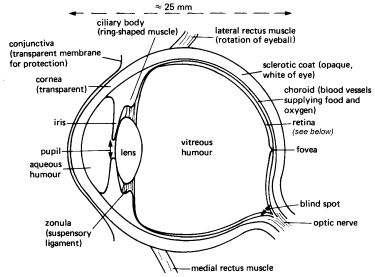
Cross sections of the eye.
Top: A horizontal section of the right eye. [Text]
Bottom: A tranverse section of the left eyeball (superior view).

- The eye has a diameter of around 25mm in an adult.
- Only one sixths of the eye's surface is exposed.
- The hole in which the eyeball rests is called the eye's orbit.
- The eye has various outer layers.
- The outermost layer of the eye consists of it's accessory structures.
Accessory Structures: Eyelashes, Eyebrows and Tears
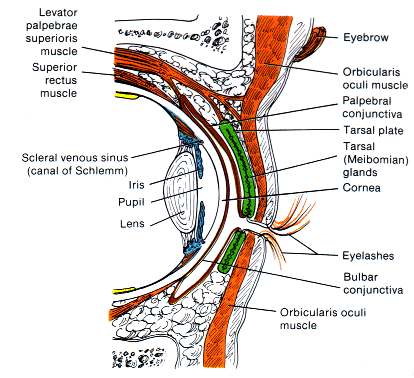
Sagittal Section Of Accessory Structures
- The eyelashes project from the border of each eyelid.
- The eyebrows arch transversely above the upper eyelids.
- The eyelashes and eyebrows together protect the eyeballs against
- Foreign Objects
- Perspiration
- Direct rays from the sun
- At the base of the hair follicles of the eyelashes are some sebaceous glands.
Sebaceous Glands
Sebaceous glands, or oil glands, with few exceptions, are connected to hair follicles. The secreting portions of the glands lie in the dermis and open into the necks of hair follicles or directly onto a skin surface (lips, glans penis, labia minora, and tarsal glands of the eyelids). Absent in the palms and soles, sebaceous glands vary in size and shape in other regions of the body. For example, they are small in most areas of the trunk and extremities, but large in the skin of the breasts, face, neck, and upper chest.
Sebaceous glands secrete an oily substance called sebum, a mixture of fats, cholesteterol, proteins, and inorganic salts. Sebum helps keep hair from drying and becoming brittle, prevents excessive evaporation of water from the skin, keeps the skin soft and pliable, and inhibits the growth of certain bacteria. When sebaceous glands of the face become enlarged because of accumulated sebum, blackheads develop. Since sebum is nutritive to certain bacteria, pimples or boils often result. The color of blackheads is due to melanin and oxidised oil, not dirt.
- The sebaceous glands at the base of the hair follicles of the eyelashes are called sebaceous ciliary glands or glands of Zeis.
- The sebaceous ciliary glands pour a lubricating fluid into the follicles.
- The lacrimal apparatus is a group of structures that produces and
drains tears.
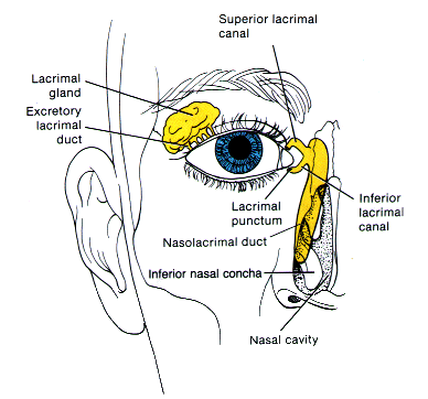
Anterior View Of Lacrimal Apparatus
- The lacrimal glands are each about the size an almond.
- The lacrimal glands secrete lacrimal fluid, or tears.
- The lacrimal glands secrete around 1ml of lacrimal fluid per day.
- Leading from the lacrimal glands are 6 to 12 excretory lacrimal ducts.
- Excretory lacrimal ducts empty tears onto the surface of the conjunctiva of the upper lid.
- The lacrimal punctum are two openings on the nasal side of the extreme edge of the eyeball.
- From the lacrimal puncta runs two ducts, called the lacrimal canals.
- There is one lacrimal canal which joins the lacrimal puncta from above, it is called the superior lacrimal canal.
- There is also one lacrimal canal which joins the lacrimal puncta from below, it is called (with great imagination) the inferior lacrimal canal.
- The lacrimal canals lead into the nasolacrimal duct.
- The nasolacrimal duct is a canal that caries the lacrimal fluid into the nasal cavity.
- There is one nasolacrimal duct on each side of the nose.
- Tears (lacrimal fluid) run from the lacrimal glands, into the excretory lacrimal ducts, onto the surface of the conjunctiva, over the surface of the eyeball, into a lacrimal puncta, from there into one of the two lacrimal canals, into the nasolacrimal duct and finally into the nasal cavity.
- Some lacrimal fluid evaporates, too.
- If an irritating substance contacts the conjunctiva, then the lacrimal glands will be stimulated to over produce lacrimal fluid.
- If the lacrimal glands over produce the lacrimal fluid, then tears accumulate.
- When tears (lacrimal fluid) accumulates on the surface of the eyeball, a situation known as watery eyes develops.
- The accumulated tears of watery eyes dilute and wash away the irritating substance.
- Watery eyes also occur when something obstructs the nasolacrimal ducts.
- Blocked nasolacrimal ducts can be caused by an inflammation of the nasal mucosa, such as a cold.
- Over production of lacrimal fluid also occurs in response to parasympathetic stimulation, caused by an emotial response.
- Over production of lacrimal fluid caused by parasympathetic stimulation can result in tears spilling over the edges of the eyelids.
- The common term for the spilling of tears over the edges of the eyelids due to over production of lacrimal fluid caused by parasympathetic stimulation is crying.
- Over production of lacrimal fluid caused by parasympathetic stimulation can even result in tears filling the nasal cavity.
- The over production of lacrimal fluid caused by parasympathetic stimulation due to an emotial response is, to our knowledge, unique to the human race.
- Lacrimal fluid is a watery solution containing:
- Salts,
- Some mucus, and
- A bacterial enzyme called lysozyme.
- The lacrimal fluid cleans the eyeball.
- The lacrimal fluid moistens the eyeball.
- The lacrimal fluid lubricates the eyeball.
- Once the lacrimal fluid runs onto the surface of the conjunctiva, it is spread medially over the surface of the eyeball by the blinking of the eyelids.
- The upper and lower eyelids are known as the palpebrae.
Eyelids (Palpebrae)
- The palpebrae shade the eyes during sleep.
- The palpebrae protect the eyes from excessive light.
- The palpebrae protect the eyes from foreign substances.
- The palpebrae spread lubricant secretions over the eyeballs.
- The palpebrae consist of (from outermost to innermost)
- the epidermis,
- the dermis,
- the subcutaneous tisue,
- fibers of the orbicularis oculi muscle,
- a tarsal plate,
- the tarsal glands (inside the tarsal plate),
- the palpebral conjunctiva (which lines the innermost part of the eyelids), and the
- the bulbar conjunctiva (which lines the outermost part of the eyeball).
- The tarsal plate is a thick fold of connective tissue.
- The tarsal plate gives form and support to the eyelids.
- Embedded inside each tarsal plate is a row of elongated modified sebaceous glands.
- The elongated modified sebaceous glands embedded inside each tarsal plate are known as tarsal or Meibomian glands.
- Tarsal glands secrete an oily substance.
- The oily substance secreted by tarsal glands helps keep the eyelids from adhering to each other.
- The conjunctiva is the innermost layer of the eyelids and the outermost layer
of the eyeball.
The Conjunctiva
- The conjunctiva is a protective mucous membrane.
- The conjunctiva is very thin.
- The conjunctiva is transparent.
- The conjunctiva consists of two parts,
- the palpebral conjunctiva (which lines the innermost part of the eyelids), and
- the bulbar (ocular) conjunctiva (which lines the outermost part of the eyeball).
- The two parts of the conjunctiva are all one long membrane, which folds onto itself.
- The next layer below the eyelids and the conjunctiva is the fibrous tunic.
The Fibrous Tunic
- The fibrous tunic is the outer coat of the eyeball.
- The fibrous tunic consists of the anterior cornea and the posterior sclera.
Tunics
Anatomically, the wall of the eyeball can be divided into three layers: fibrous tunic, vascular tunic or urea, and retina or nervous tunic.
- The outer surface of the cornea is covered by an epithelial layer.
- The epithelial layer which covers the outer surface of the cornea is continuous with the epithelium of the bulbar conjunctiva.
- The cornea is a fibrous coat.
The Cornea
- The cornea is a relatively thick layer.
- The cornea is nonvascular.
- The cornea is transparent.
- The cornea has a refractive index of 1.38.
- The cornea is curved.
- The cornea helps focus light.
- Around 75% of the focusing, or refracting, occurs at the cornea.
- The cornea is connected directly to the sclerotic coat.
Sclerotic Coat
- The sclerotic coat is opaque.
- The sclerotic coat is white.
- The sclerotic coat extends all the way round the back of the eye, until it touches the cornea.
- The sclerotic coat is also called the sclera.
skleros = hard
- The sclera is a coat of dense connective tissue.
- The sclera gives shape to the eyeball.
- The sclera makes the eyeball more rigid.
- The sclera protects it's inner parts.
- The posterior surface of the sclera is pierced by the optic foramen.
Optic Foramen
- The optic foramen encircles the optic nerve.
- Below the fibrous tunic is the vascular tunic or urea.
Vascular Tunic or Urea
- The vascular tunic consists of three portions.
- The choroid,
- The ciliary body, and
- The iris.
vascular = containing (or pertaining to) many blood vessels
- Lining most of the internal surface of the sclera is the choroid.
- The choroid is highly vascularized.
Choroid
- The choroid provides nutrients to the posterior surface of the retina.
- The choroid has a brown/black appearance.
- The brown/black appearance of the choroid is due to the melanocytes.
- Melanocytes are pigmented cells that synthesize melanin.
Melanocytes
- Melanocytes are located between and beneath cells in the choroid.
- In the part of the vascular tunic near the lens, the choroid becomes the ciliary body.
Ciliary Body
- The ciliary body
- Unsorted Past This Point
- At the junction of the sclera and cornea is an opening knows as the scleral venous sinus.
- The scleral venous sinus is also called the canal of Schlemm.
- The eye is filled with fluid.
- The fluid in the eye maintains the even spherical shape of the eye wall.
- The fluid is made inside the ciliary body.
- The ciliary body is also made up of muscles.
- The ciliary body allows the eye to focus at different distances.
- The eye is divided into two chambers by the lens.
The Lens
- The lens is is transparent.
- If the lens becomes cloudy or hazy, it is called a cataract.
- Cataract are common in older individuals.
- The lens is elastic.
- The lens is biconvex.
- The lens is not homogenous.
- The lens consists of successive fibrous layers.
- The fibrous layers of the lens are gradually added throughout life.
- The lens becomes harder over time.
- The lens has a refractive index which varies from 1.42 at the centre to about 1.38 at the periphery.
- The retina is the light-sensitive part of the eye.
The Retina
- The retina is also layered.

Structure of the Retina
- The layers of the retina, from the outermost to innermost, are, amongst others,
- the inner limiting membrane,
- the nerve cells (or ganglion cells),
- the relay nerve cells (or bipolar cells),
- the rods and cones, and finally
- the melanin.
- The rods and cones are the retina's light-sensitive receptors.
Rods and Cones
- Rods are slender rod-like elements.
- Cones are narrow conical elements.
- Cones are the receptors of most distinct vision.
- Rods and cones have diameters in the range of microns (10-6 m).
- There are roughly twenty times more rods than there are cones.
- The rods dominate the retinal periphery.
- The cones dominate the optical axis.
- Rods do not register colour.
- Rods are not equally sensitive to all wavelengths.
- Rods display the maximum absorption in the green region of the spectrum (500×10-9 — 580×10-9m), at about 510×10-9m.
- Cones descriminate colour.
- There are three types of cones: red-sensitive cones , green-sensitive cones and blue-sensitive cones.
- Rods operate at low intensities.
- Cones are stimulated only by fairly high light intensities.
- Rods are responsible for vision in dim light (night and twilight, or scotopic vision).
- Cones are responsible for the more acute vision of daylight (colour or phototopic vision).
Scotopic Vision
Rods contain a light sensitive pigment called rhodospin, which contains a protein called scotopsin and a variant of the pigment called retinene.
Photopic Vision
The different cone types contain different light sensitive pigments, but they are all thought to consist of a protein called photopsin and variants of the pigment retinene.
- Rods and cones contain a light sensitive pigment.
- Rods use the pigment rhodospin.
- Rhodopsin is reddish-purple.
- Rhodopsin is sometimes called
visual purple
. - Red-sensitive cones use the pigment erythrolabe.
- Green-sensitive cones use the pigment chlorolabe.
- Blue-sensitive cones use the pigment cyanolabe.
- Rhodospin, erythrolabe, chlorolabe and cyanolabe all contain variants on the pigment retinene.
- Retinene is chemically similar to Vitamin A.
- Vitamin A is also called retinol.
- When rhodospin is exposed to light, it bleaches.
- Rhodospin first bleaches yellow, then clear.
- The bleaching of the rhodospin stimulates the rod cell.
- When the rod cell is stimulated, it in turn stimulates it's associated nerve cells.
- Bleached rhodospin is rapidly restored to it's original condition by an enzymatic process.
- The enzymatic process uses Vitamin A derived from the blood supply.
- The blood supply comes from the choroid.
- The choroid is two layers below the rods and cones.
- Similar processes occur with the cones and their erythrolabe, chlorolabe and cyanolabe pigments.
- Several rods share the same neural pathways to the brain.
- Cones have less sharing of nerve fibres.
- Cones have very little sharing of nerve fibres in the foveal region.
The Fovea
- The fovea is the region of most acute vision.
- The fovea contains only cones.
- In the fovea, the nerve fibre are displaced radially away.
- Radially displaced fibres allow light to strike the cones directly.
- When the fibres are not radially displaced the photons must first traverse the other layers of the retina.
- The fovea is a small depression.
- The fovea is in the centre of the yellow spot.
The Yellow Spot or Macula
- The yellow spot is also called the macula.
- The yellow spot is situated on the optical axis.
- The yellow spot is in the centre of the retina.
- The yellow spot is
yellowish
. - The yellow spot has a diameter of about 1mm.
- The blind spot is a creamy white spot.
The Blind Spot
- There are no receptors in the blind spot.
- When using one eye only, the blind spot causes a blind field
- The blind field is located about 6° horizontally and 8° vertically.
- The blind spot is located about 10° from the optical axis on the nasal side.
- The blind spot is located where the optical nerve leaves the eye.
Optic Nerve
- There are two optical nerves: one for each eye.
- There are about 1.1 million nerve fibres in each optic nerve.
- The optic nerve is much akin to brain tissue.
Mechanisms of the eye
- Accommodation is one of the focusing mechanisms of the eye.
Accommodation
- In order to focus rays from objects at varying distances, the lens must change it's refractive power.
- To change it's refractive power, the lens changes shape.
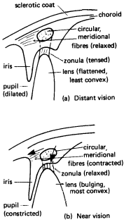
- The exact shape of the lens is determined by seventy or so suspensory ligaments.
- The suspensory ligaments attached to the lens are called zonula.
- The zonula are attached radially around the lens.
- The zonula pull the edges of the lens towards the clilary body.
- When the eye is accommodated for distant vision, both the circular and meridional fibres of the ciliary muscle are relaxed. (a)
- When both the circular and meridional fibres of the ciliary muscle are relaxed, the zonula is stretched.
- When the zonula is stretched it pulls the elastic lens into a flattened shape.
- When the eye is accommodated for near vision, both the circular and meridional fibres of the ciliary muscle contract. (b)
- When both the circular and meridional fibres of the ciliary muscle contract, the tension in the zonula is released.
- When the ciliary muscle contracts, the ciliary process and choroid move forward toward the lens.
- When the tension in the zonula is released, it allows the the elastic lens to bulge.
- The lens of the eye is elastic.
- Because the lens is elastic, it bulges, shortens, and thickens.
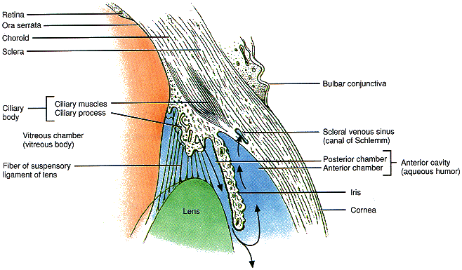
Close Up Of Sagittal Section Of Accessory Structures
- The ciliary process is the aqueous humor factory.
- The aqueous humour is drained out of the scleral venous sinus.
The maths of the eye
- Refractive Power
The power of a refracting surface is defined as
power = 1
focal lengthThe units of focal length are metres (m), therefore the units of refractive power are per meters (m-1). There is another name for the units of refractive power though, they are called dioptres (D).
The power of the unaccommodated eye is approximately 59 D.
The relationship between u, the distance between the object and the lens, v, the distance between the virtual image and the lens, and f, the focal length of the lens, is
1
u+ 1
v= 1
fWhen using this equation one must follow the sign convention, which states
A real object or image distance is positive.
A virtual object or image distance is negative.
The diseases of the eye
- Infection of the tarsal glands produces a tumour or cyst on the eyelid called a chalazion.
- When the blood vessels of the bulbar conjunctiva are dilated and congested due to local irritation or infection, the person has bloodshot eyes.
- Infection of the sebaceous ciliary glands is called a sty.
- The term astigmatism describes the condition wherein
the eye's reflecting surfaces have an uneven curvature.
Astigmatism
- It is typically the cornea which has the uneven surface causing astigmatism.
- When an eye is afflicted with astigmatism, it can focus rays in some planes better than others.
- The condition known as astigmatism may be corrected using a suitable cylindrical lens.
- These lenses are called toric or cylinder lens.
Sample Question
A young person and an older person each wear spectacles with converging lenses to view objects nearer than 1.0m to correct different problems caused by defects in their vision.
State the causes of the defects in each case. Explain how the defects give rise to the problems.
Sample Answer
Clinical Treatments
- Corneal Transplants Corneal Transplants
- Corneal transplants are the most common organ transplant operation.
- Corneal transplants are the most successful type of transplant, too.
- Rejection of a transplanted cornea is rare.
- Because the cornea is avascular, antibodies that might cause rejection do not circulate there.
- During the operation, the defective cornea is first removed, and then a donor cornea of similar diameter is sewn in.
- There is a shortage of donor cornea.
- The shortage of donated corneas has been partially overcome by the development of artificial corneas made of plastic.
Bibliography
Medical Physics, second edition, by Jean A Pope (Options on Physics series).
Principles of Anatomy and Physiology, seventh edition, by Gerard J. Tortora and Sandra Reynolds Grabowski. ISBN 0-06-046702-7.
The Eye Clinic, http://ofcn.org/cyber.serv/hwp/hwc/eye/information/
Some diagrams are almost direct copies (with some minor changes to improve presentation) of those found in the above mentioned sources.
